


Abstract
The work concerns studying electrophysiological parameters and, based on histological studies, morphological parameters of the retina in different age groups of Campbell rats with Retinitis Pigmentosa (RP) before and after Retinalamin treatment. 50 Campbell rats, as an experimental model of inherited RP, were used in the study. Rats were divided into two age groups, with 25 rats in each. Group I consisted of newborns up to 3 weeks, and Group II consisted of adults aged 3.5 ± 0.3 months. Retinalamin injections were given parabulbarly to all animals for 10 days. The total bioelectrical activity of the a, b, and c waves and the average value of the b-wave amplitude were studied using the electroretinography (ERG) method.
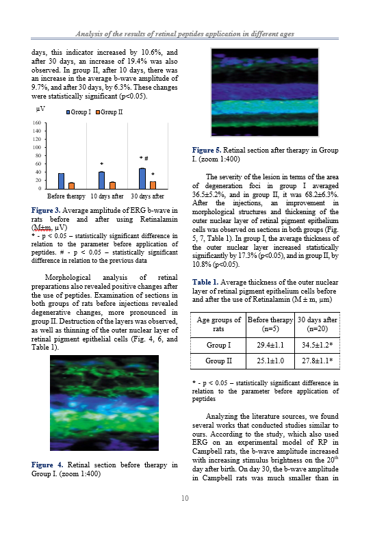
Also, the thickness of the outer nuclear layer of retinal pigment epithelium cells was studied in a histological examination. Studies were conducted before the start of Retinalamin injections and in dynamics after 10 and 30 days.
According to the ERG results, a decrease in the total bioelectrical activity of the rat retina and the average b-wave amplitude were already observed in 19-day-old rats. A statistically significant increase in these indicators was observed in both groups after 10 and 30 days of Retinalamin injections. Histological analysis of retinal preparations also revealed positive changes after the peptide administrations. An improvement in morphological structures and thickening of the outer nuclear layer of retinal pigment epithelial cells were noted in both groups. More significant positive dynamics were in Group I of newborn rats.
Conclusion. Retinalamin treatment in newborn and adult Campbell rats with retinitis pigmentosa stabilized the processes developing in the retina. At the same time, the maximum positive effect was manifested in newborn rats.
References
Bhootada Y, Kotla P, Zolotukhin S, Gorbatyuk O, Bebok Z, Athar M, Gorbatyuk M. Limited ATF4 Expression in Degenerating Retinas with Ongoing ER Stress Promotes Photoreceptor Survival in a Mouse Model of Autosomal Dominant Retinitis Pigmentosa. PLoS One. 2016 May 4;11(5):e0154779. https://doi.org/10.1371/journal.pone.0154779
Nagy D, Schönfisch B, Zrenner E, Jägle H. Long-term follow-up of retinitis pigmentosa patients with multifocal electroretinography. Invest Ophthalmol Vis Sci. 2008 Oct;49(10):4664-71. https://doi.org/10.1167/iovs.07-1360.
Fahim A. Retinitis pigmentosa: recent advances and future directions in diagnosis and management. Curr Opin Pediatr. 2018 Dec;30(6):725-733. https://doi.org/10.1097/MOP.0000000000000690.
Kharauzov A.K., Etingof R.N. Change on schroedinger rats line Campbell in the development of hereditary retinal degeneration. Physiology (Bethesda). 2014; 9:65-71.
Khasanova NH, Belyaeva AV. Results of Retinalamin usage in retinal diseases. Russian Journal of Clinical Ophthalmology, 2008;9(3):77-82. Russian.
Khavinson VKh, Proniaeva VE, Lin'kova NS, Trofimova SV, Umnpv RS. [Molecular-physiological aspects of peptide regulation of function of the retina in retinitis pigmentosa]. Fiziol Cheloveka. 2014 Jan-Feb;40(1):129-34. Russian.
Liu W, Liu S, Li P, Yao K. Retinitis Pigmentosa: Progress in Molecular Pathology and Biotherapeutical Strategies. Int J Mol Sci. 2022 Apr 28;23(9):4883. https://doi.org/10.3390/ijms23094883.
Miliushina LA, Kuznetsova AV, Aleksandrova MA. [Experimental models of human retinal degenerations: genetic models]. Vestn Oftalmol. 2013 Mar-Apr;129(2):81-5. Russian.
Nuri NH. Dynamics of the course of Retinitis Pigmentosa in a child with macular involvement over 6 years. Ophthalmology Cases & Hypotheses. 2021 Mar 1;2(1):1-4. https://doi.org/10.30546/2788-516X.2021.2.1.1.
Pan M, Yin Y, Wang X, Wang Q, Zhang L, Hu H, Wang C. Mice deficient in UXT exhibit retinitis pigmentosa-like features via aberrant autophagy activation. Autophagy. 2021 Aug;17(8):1873-1888. https://doi.org/10.1080/15548627.2020.1796015.
Takhchidy KhP, Gavrilova NA, Komova OYu et al. [Influence of stem/progenitor cells on functional status and level of degenerative changes of the retina in Campbell rats]. Ophthalmosurgery. 2010;3:33-38. Russian.
Trofimova SB, Khludieva TA, Ivko OM, Anas AD. [The effect of the bioregulating therapy on the quality of life of elderly patients with retinal pathology]. Adv Gerontol. 2006;18:96-9. Russian.
Zhang Q. Retinitis Pigmentosa: Progress and Perspective. Asia Pac J Ophthalmol (Phila). 2016 Jul-Aug;5(4):265-71. https://doi.org/10.1097/APO.0000000000000227.
Zolnikova IV, Rogatina EV, Egorova IV, Zueva MV. Multifocal ERG, chromatic macular ERG and retinocortical time in congenital stationary night blindness. Abstracts of 48th ISCEV Symposium, Fremantle, Australia, 2010. - р.86.

This work is licensed under a Creative Commons Attribution 4.0 International License.
Copyright (c) 2023 Azerbaijan Journal of Physiology